
Great outcomes depend on great technology — but technology only delivers when paired with the experience to use it well. Every instrument in this practice has been chosen to give patients the most accurate, safest result possible.
The gold-standard optical biometer — it measures the eye with exceptional precision to determine exactly which lens power will give the sharpest vision after cataract surgery. Used for every patient before surgery.

The world's most validated formula for planning toric (astigmatism-correcting) lenses. It accounts for the full contribution of the cornea to astigmatism — something older methods miss — producing consistently more accurate outcomes.
A sophisticated instrument that maps the unique optical characteristics of each patient's eye, separating corneal from internal aberrations. This allows Dr. Scheepers to fine-tune lens selection beyond what standard measurements provide — particularly valuable for patients with prior laser eye surgery or unusual corneas.

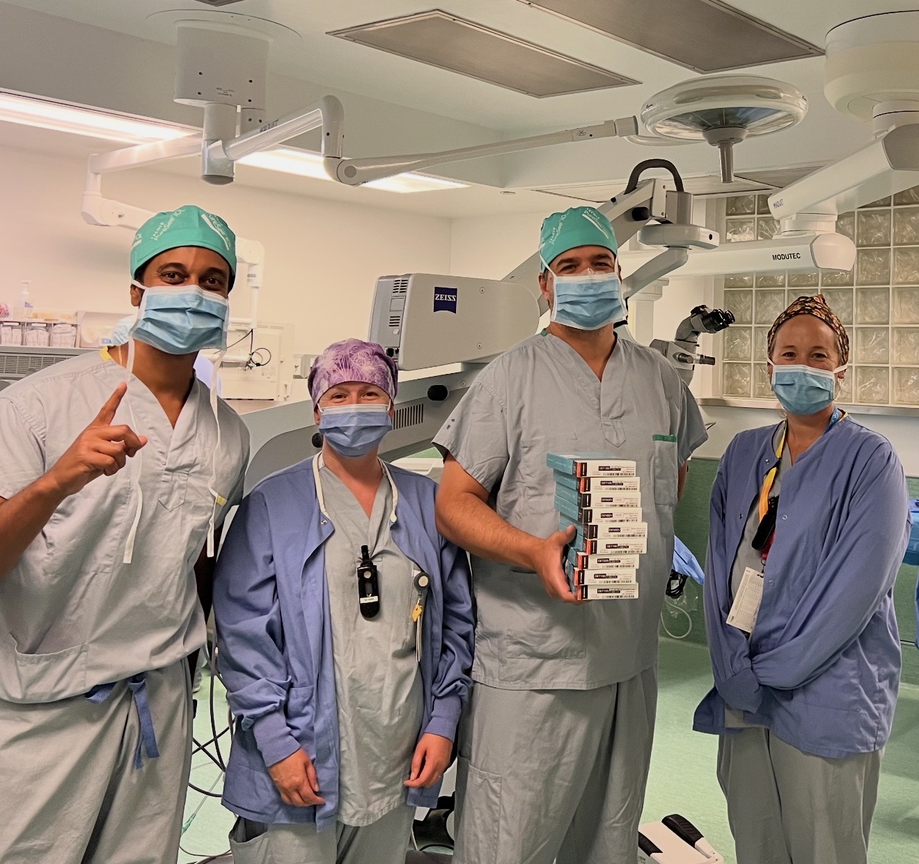
The Callisto projects a real-time digital overlay onto the surgical microscope during the operation, guiding every step of the procedure with computer precision — no ink marks, no estimation. The images below are from real surgeries performed by Dr. Scheepers.
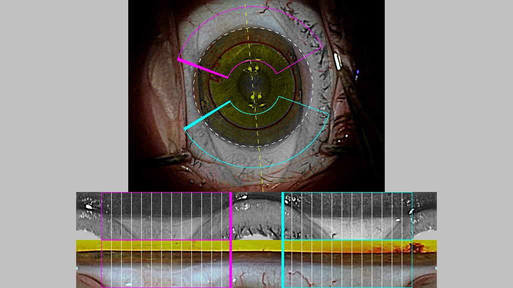
Before surgery, the Callisto captures the eye's unique landmarks. This reference guides every overlay during the operation, regardless of head position.
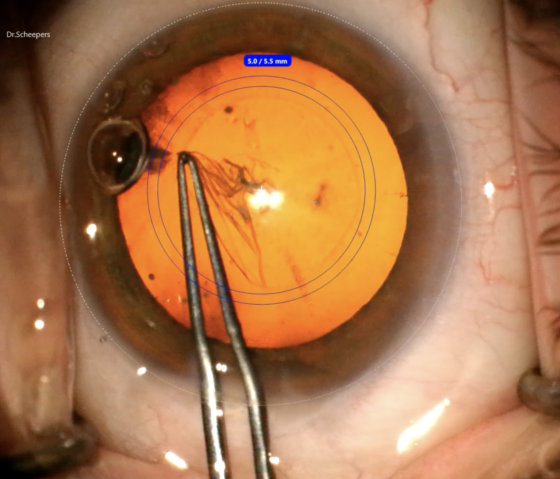
A circular overlay centred precisely on the visual axis guides the creation of the capsular opening — critical for optimal lens centration, especially with premium implants.
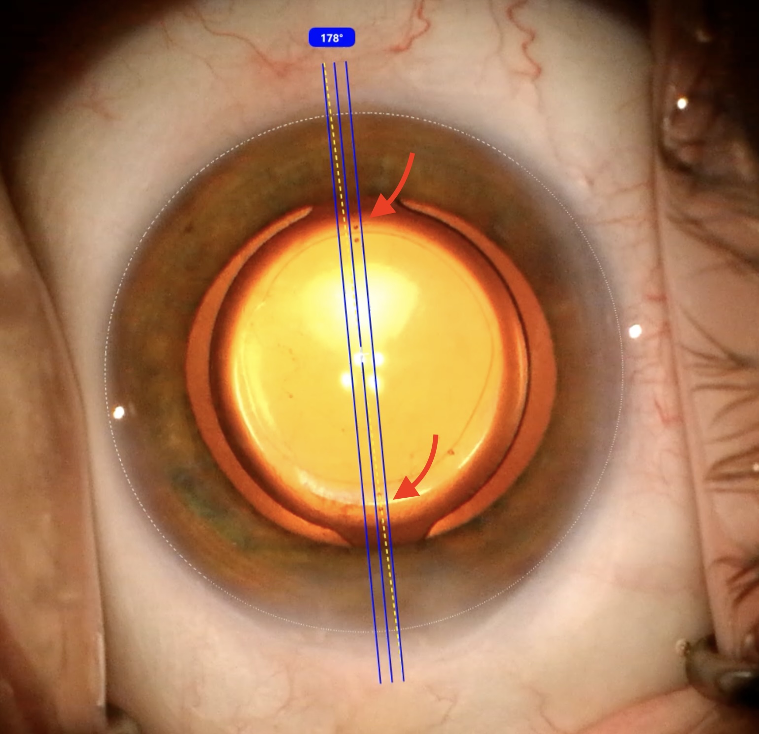
For astigmatism correction, the Callisto projects the exact target axis onto the microscope. The lens is rotated until its alignment marks (shown by red arrows) match the digital overlay — precise to within a degree, with no ink required.
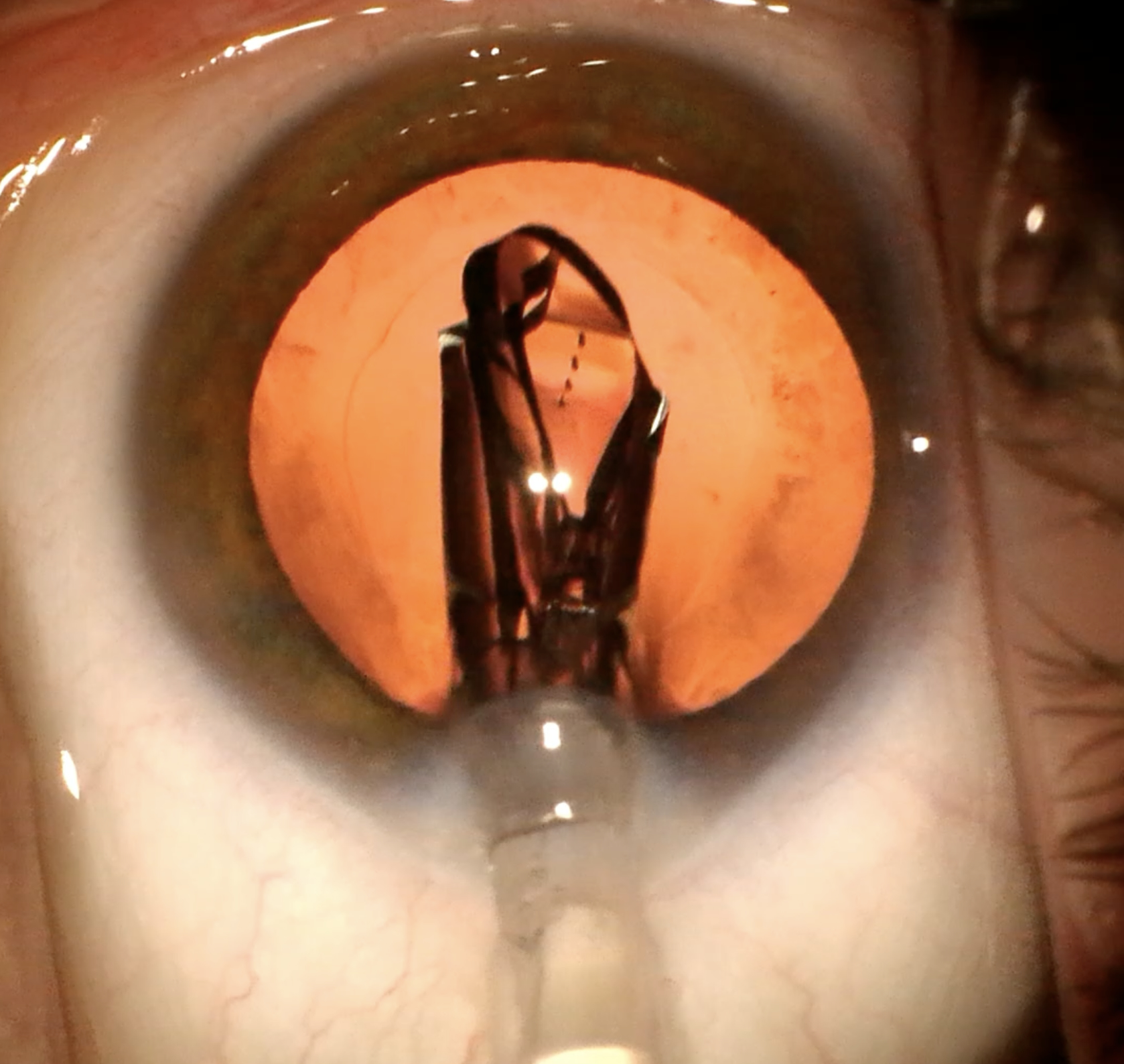
In over 99% of cases, the intraocular lens arrives preloaded in a sterile cartridge and is never manually handled. It passes directly from cartridge into the eye through a small, self-sealing incision.
In 2024, Dr. Scheepers became the first surgeon in BC to implant the TECNIS PureSee (Johnson & Johnson) — a next-generation extended depth-of-focus lens offering a natural, seamless range of vision with excellent low-light quality.
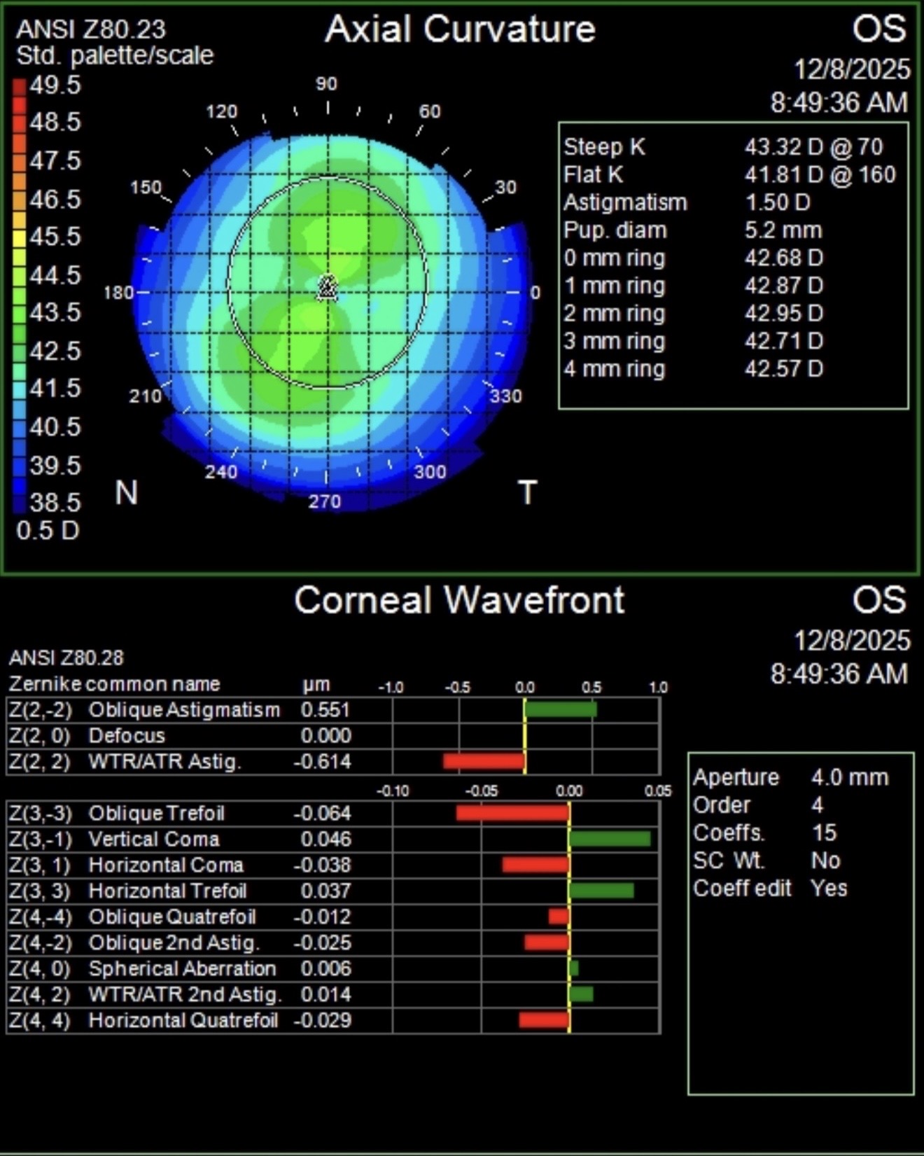
Dr. Scheepers uses the iTrace and ZEISS Atlas corneal topographers across his clinics. These instruments create detailed colour maps of the corneal surface — essential for planning astigmatism correction at the time of cataract surgery, whether through a toric lens or through small astigmatic keratotomy incisions made during the procedure itself.
The before-and-after maps below show a real patient example. Pre-operatively, the cornea showed 1.50 dioptres of astigmatism (the classic bow-tie pattern). Following astigmatic keratotomy incisions at the time of cataract surgery, astigmatism was reduced to just 0.14 dioptres — a near-spherical result.
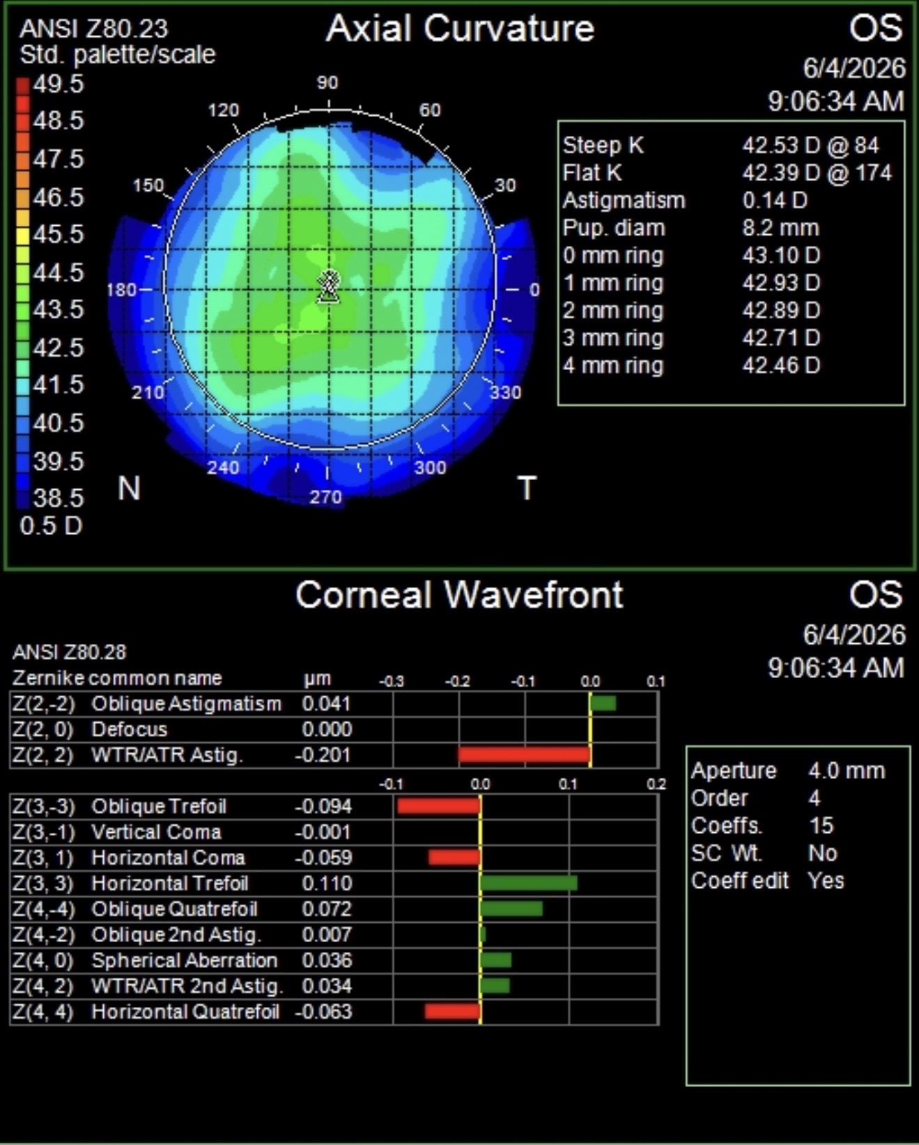
Small relaxing incisions placed in the corneal periphery at the time of cataract surgery can meaningfully reduce astigmatism at no additional lens cost. Topography guides the planning, and post-operative maps confirm the result.
High-resolution retinal and optic nerve imaging available on-site — used for monitoring macular degeneration, diabetic eye disease, glaucoma, and other retinal conditions at every visit.

On-site automated perimetry for glaucoma monitoring, visual pathway assessment, and neuro-ophthalmology — results available at your consultation, not weeks later.